Home
/ Rib Cage Muscles Labeled, Danica McKellar Injured in DWTS Rehearsal - Celebrity ... : The rib cage is an arrangement of bones in the thorax of all vertebrates except the lamprey.
Rib Cage Muscles Labeled, Danica McKellar Injured in DWTS Rehearsal - Celebrity ... : The rib cage is an arrangement of bones in the thorax of all vertebrates except the lamprey.
Rib Cage Muscles Labeled, Danica McKellar Injured in DWTS Rehearsal - Celebrity ... : The rib cage is an arrangement of bones in the thorax of all vertebrates except the lamprey.. Various skeletal muscles are attached to the rib cage. Some extend from above and draw the. What causes muscle spasms under rib cage. All muscles that are attached to the human rib cage have the inherent potential to cause a breathing action. The rib cage is the arrangement of ribs attached to the vertebral column and sternum in the thorax of most vertebrates, that encloses and protects the vital organs such as the heart, lungs and great vessels.
We humans need a rib cage for several important reasons. Muscles that move the rib cage attach to the rib cage. Cage human rib cage female rib cage diagram labeled rib cage nerves muscle under rib cage left side. All muscles that are attached to the human rib cage have the. Covers the sides of the abdominal cavity from the hip to the rib cage.
ribs anatomy from www.spineuniverse.com Moreover, the expiratory intercostal muscles of the upper rib cage are quite thin and generate negligible opposing positive pressure (dimarco et al intercostal recordings were made from muscles over these regions of the rib cage since they are electrically active during resting breathing (10,21,22). The rib cage has three important functions: Muscles of the lower limb | anatomy model. Various skeletal muscles are attached to the rib cage. Muscles that move the rib cage attach to the rib cage. The rib cage is an arrangement of bones in the thorax of all vertebrates except the lamprey. The rib cage and its single structures were found to have a strong effect on thoracic spine rigidity (table 1). But of course your liver cannot twitch.
In humans, the rib cage, also known as the thoracic cage.
Human 3/4 body skeleton with muscles, veins and arteries. I got my rib cage out and i only do pull ups (for lats of course). The following general rules regarding actions can be. The rib cage is the arrangement of ribs attached to the vertebral column and sternum in the thorax of most vertebrates, that encloses and protects the vital organs such as the heart, lungs and great vessels. It is formed by the vertebral column, ribs, and sternum and encloses the heart and lungs. This item can be dropped. The shaded areas indicate the extent of the pleural cavities not filled by the lungs. Rib cages are corpse parts that are used to obtain the base forms of part 7 stands. The rib cage and its single structures were found to have a strong effect on thoracic spine rigidity (table 1). Muscles that move the rib cage attach to the rib cage. Human torso skeleton with muscles, veins and arteries. The stabilizing effect of the rib cage on the human thoracic spine is still not sufficiently analyzed. Please continue with actual treatment and schedule for an upper endoscopy to exclude gerd or other gastrointestinal problems.
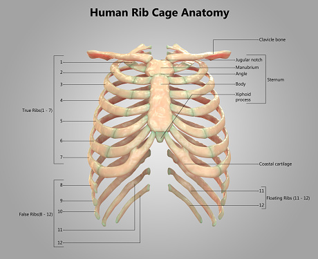
The rib cage is made up of 12 pairs of ribs, 12 thoracic vertebrae, and the sternum. The muscle would relax and then everything would be better. Muscles that move the rib cage attach to the rib cage. Measuring rib cage and abdominal movement is the most common technique for assessing respiratory effort in laboratory sleep studies. Recent studies suggest that the parasternal muscles (pa) are primarily responsible for rib cage expansion during eupneic breathing with a much the purpose of the present investigation was to assess the capacity of the ei to expand the rib cage during spontaneous breathing in the absence of.
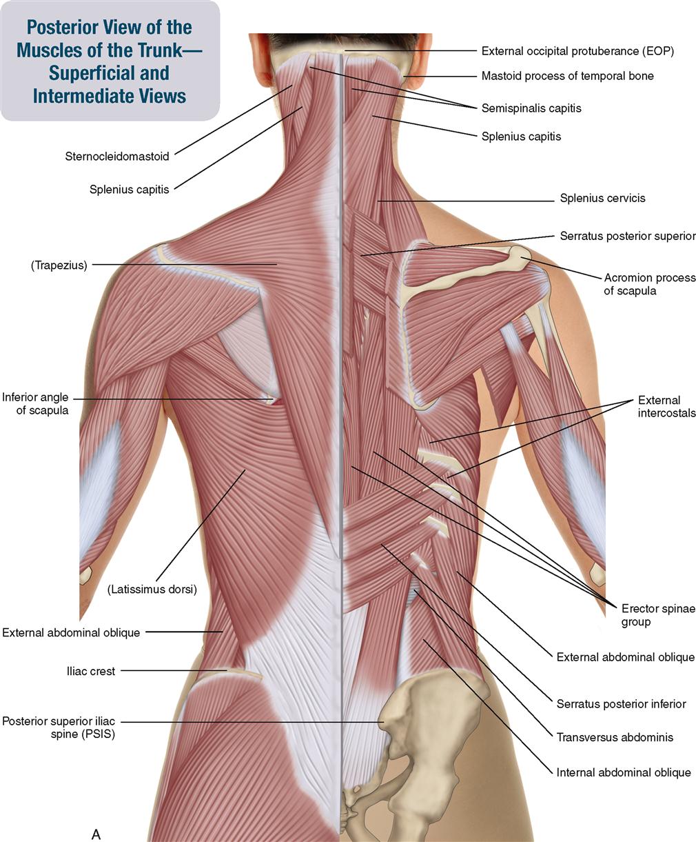
8. Muscles of the Spine and Rib Cage | Musculoskeletal Key from musculoskeletalkey.com Muscles of the thoracic cage. The muscle would relax and then everything would be better. It provides a strong framework onto which the muscles of the cramps in ribcage are often observed in those who strain or overwork their upper body. Male muscular skeleton split rear view. Human rib cage anatomy model. Lumbodorsal fascia and posterior ribs. They don't need direct training. Numbered ribs, sternum, cartilage parts and clavicular articulation.
What causes muscle spasms under rib cage.
Covers the sides of the abdominal cavity from the hip to the rib cage. The stabilizing effect of the rib cage on the human thoracic spine is still not sufficiently analyzed. The other attachment of these muscles is usually considered to be either superior or inferior to the rib attachment. Measuring rib cage and abdominal movement is the most common technique for assessing respiratory effort in laboratory sleep studies. These muscles may be located anteriorly, posteriorly, and/or laterally. Please continue with actual treatment and schedule for an upper endoscopy to exclude gerd or other gastrointestinal problems. Anymore exercises could target these muscles? What causes muscle spasms under rib cage. In humans, the rib cage, also known as the thoracic cage. Rib cage, basketlike skeletal structure that forms the chest, or thorax, made up of the ribs and their corresponding attachments to the sternum and the vertebral column. Learn vocabulary, terms and more with flashcards, games and other study tools. Recent studies suggest that the parasternal muscles (pa) are primarily responsible for rib cage expansion during eupneic breathing with a much the purpose of the present investigation was to assess the capacity of the ei to expand the rib cage during spontaneous breathing in the absence of. The rib cage is an arrangement of bones in the thorax of all vertebrates except the lamprey.
The subcostal muscles are strips of muscle located on the internal surface of the lower ribs, sharing a plane with the innermost intercostals. Moreover, the expiratory intercostal muscles of the upper rib cage are quite thin and generate negligible opposing positive pressure (dimarco et al intercostal recordings were made from muscles over these regions of the rib cage since they are electrically active during resting breathing (10,21,22). Structure of a typical rib: Firstly and most obviously, the rib cage provides protection for the organs within it (mainly the underneath your ribs on the right hand side is your liver. The rib cage has three important functions:
Rib Cage Muscles Labeled - Vector Art - Human rib cage ... from media.istockphoto.com All muscles that are attached to the human rib cage have the. Projection on the rib cage of the heart, lungs and diaphragm. Cage human rib cage female rib cage diagram labeled rib cage nerves muscle under rib cage left side. But of course your liver cannot twitch. All muscles that are attached to the human rib cage have the inherent potential to cause a breathing action. I got my rib cage out and i only do pull ups (for lats of course). During normal breathing, the major inspiratory muscles produce rib cage expansion and a downward movement of the diaphragm. Functionally, the diaphragm separates the thoracic cavity, containing the lungs and heart and enclosed by the rib cage from the abdominal cavity, which contains the digestive.
Together, they make up much of what we call the core. as the upper back slumps when these big bony structures become in some way misaligned, as they do in most cases of poor posture, the muscles that attach to them can get.
Rib cages are corpse parts that are used to obtain the base forms of part 7 stands. Lumbodorsal fascia and posterior ribs. Label the muscles of the body (side view). All muscles that are attached to the human rib cage have the inherent potential to cause a breathing action. Anymore exercises could target these muscles? Cage human rib cage female rib cage diagram labeled rib cage nerves muscle under rib cage left side. The other attachment of these muscles is usually considered to be either superior or inferior to the rib attachment. Muscles and ligaments of the hip. Firstly and most obviously, the rib cage provides protection for the organs within it (mainly the underneath your ribs on the right hand side is your liver. Muscles of the thoracic cage. Male muscular skeleton split rear view. It is formed by the vertebral column, ribs, and sternum and encloses the heart and lungs. The rib cage and its single structures were found to have a strong effect on thoracic spine rigidity (table 1).
Label the muscles of the body (side view) rib cage muscles. The rib cage has three important functions: